Freehand Elastography – Breast Imaging
JC Bamber, C Uff, L Garcia, J Fromageau, NL Bush; in collaboration with D Napolitano, G McLaughlin, Zonare Medical Systems Inc.; S Allen, L Hastings, Radiology Department; A Gee, G Treece and R Prager, Engineering Department, University of Cambridge; E Brusseau, Inserm unité 630, Université de Lyon, France.
Source of funding: EPSRC, Zonare Medical Systems Inc.
Freehand elasticity images, created by using ultrasound to measure the internal tissue strain that results from external palpation, have been shown to provide useful information in various clinical applications, including the differential diagnosis of breast lesions.
A real-time implementation of our 2D cross-correlation strain imaging algorithm using I/Q data within Zonare Medical Systems’ Z.one™ ultrasound scanner has demonstrated excellent spatial resolution and strain signal-to-noise ratio that extends well into the low strain region, making it suitable for strain imaging using internal vascular pulsations, as an alternative to applying probe pressure.
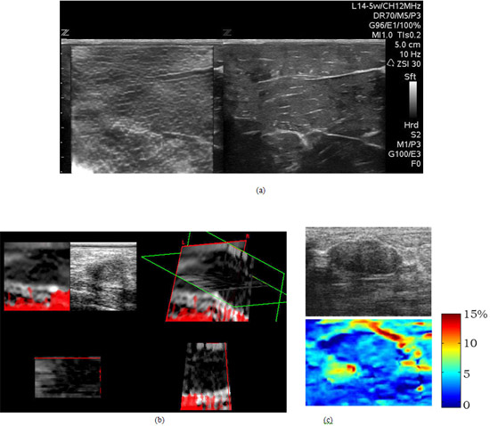
The high resolution nature of these images is particularly apparent in the phantom image shown in Figure 2a. With A Gee and colleagues at the Engineering Department University of Cambridge, 3D axial strain imaging has been implemented using a GE mechanically-swept 4D 7.5MHz probe attached to a Dynamic Imaging Diasus™ ultrasound scanner (Figure 2b). In collaboration with E Brusseau at the University of Lyon we are investigating the benefits, for breast tumour imaging, of a novel 2D locally-regularized strain estimation algorithm, which provides high quality elastograms when used with very high strains (Figure 2c).
 Fig. 2. (a) Frame from a sequence obtained using the Z.one™ real-time elastography feature that shows the ability to visualize fine stiffness detail (left) in a turkey breast phantom (B-mode image on the right). (b) a 3D elastogram of a breast tumour that looks characteristically larger on elastography than on conventional echography and contains a central very stiff region. Three orthogonal slices through the 3D strain dataset are shown top-left, bottom-left and bottom-right, while the top-right images shows all three slices placed in their correct spatial relationships and viewed from a user-selected direction. The red mask over parts of the images indicates where reliability of the strain estimates is judged to be poor. (c) Breast fibroadenoma axial strain image obtained with a novel 2D regularized strain estimator showing apparently good quality even for high (15%) strains.