Development of ultrasound motion estimation techniques to optimise radiation therapy delivery
Tuathan O’Shea, a member of the Imaging for Radiotherapy Adaptation team is researching novel ways to use imaging techniques to improve radiation therapy delivery.
It is well known that for many anatomical sites the tumour remains mobile during radiation therapy (RT) requiring margins around the target to account for motion (known as intra-fraction motion). With the application of imaging and improved information on tumour position, margins can be decreased, reducing radiation to surrounding healthy tissue and potentially allowing increased tumour dosage.
The focus of the research is using ultrasound imaging for this purpose. Ultimately, this will be achieved by imaging and accounting for internal tissue motion in “real-time”, through methods such as (a) arresting motion, (b) gating or (c) tracking tissue motion.
Ultrasound holds a number of advantages over conventional RT imaging methods including (i) producing no ionising radiation, (ii) being non-invasive and (iii) achieving fast imaging rates. To date research has focused on investigating the use of ultrasonic speckle to estimate tissue motion. At adequate imaging rates, the characteristics of speckle mean that tissue can be tracked even in the absence of anatomical features. A study was performed to look at the accuracy of prostate speckle tracking compared to kV x-ray imaging on the state-of-the-art CyberKnife® system. Current studies include development of methods for robust estimation of liver feature (i.e. blood vessels) motion and collaborative work on using ultrasound monitoring of breath-hold and a Monte Carlo investigation of the dosimetric impact of the ultrasound probe. A clinical study using with Clarity®, the first commercial ultrasound intra-fraction motion monitoring device is underway.

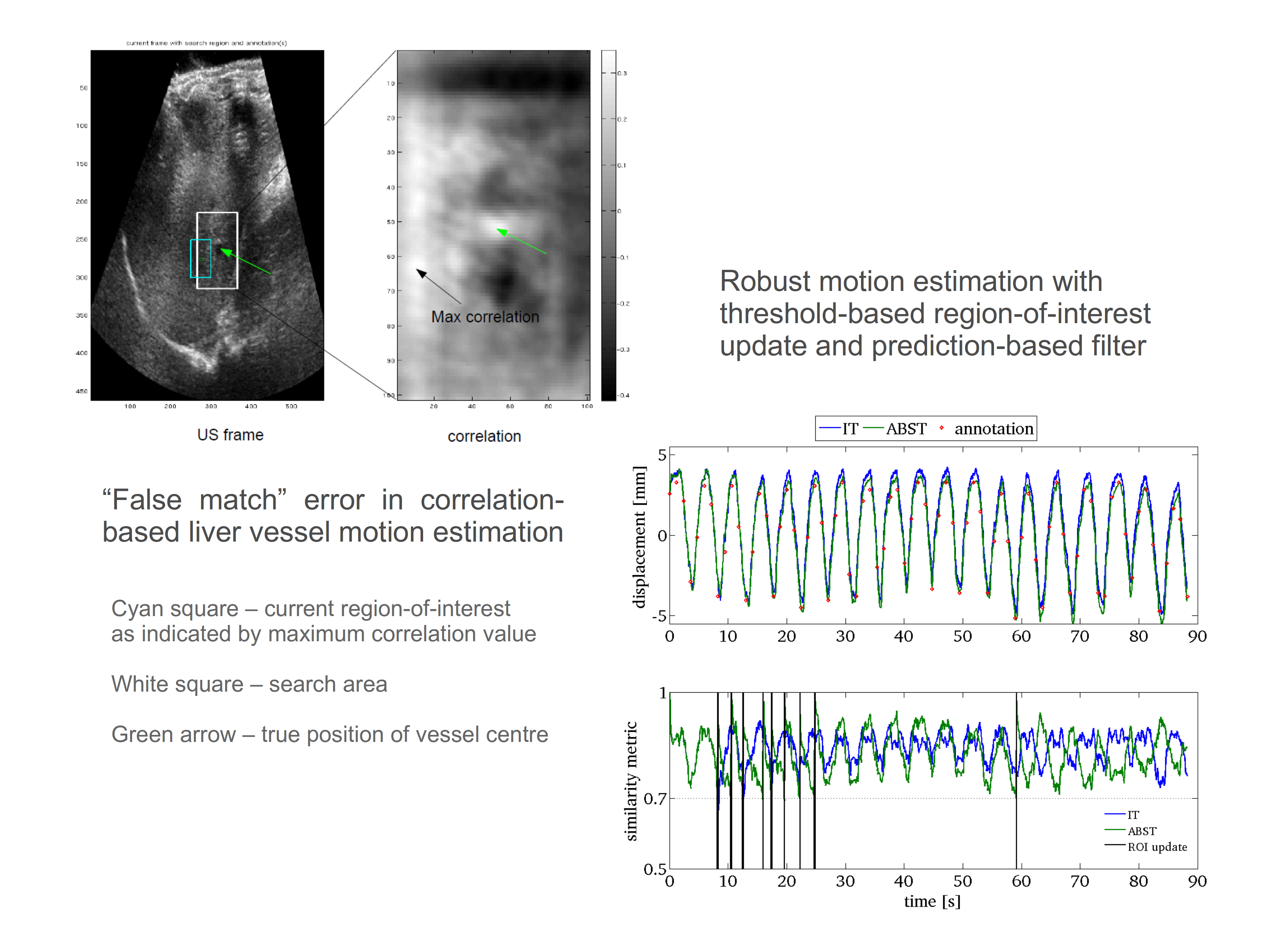
Figure 1. Robust correlation-based liver vessel motion estimation.
Robust motion estimation
For tumour tracking, the ultrasound-based motion estimate must remain accurate during the radiation beam-on time. Changes in tissue appearance can present challenges for motion estimation algorithms over long imaging periods. Current research is investigating methods for robust tracking of liver vessel motion due to cardiac and respiratory motion in long (2 to 5 minute) ultrasound sequences. Tracking methods under investigation include regularised normalised cross-correlation-based template matching and optical flow.

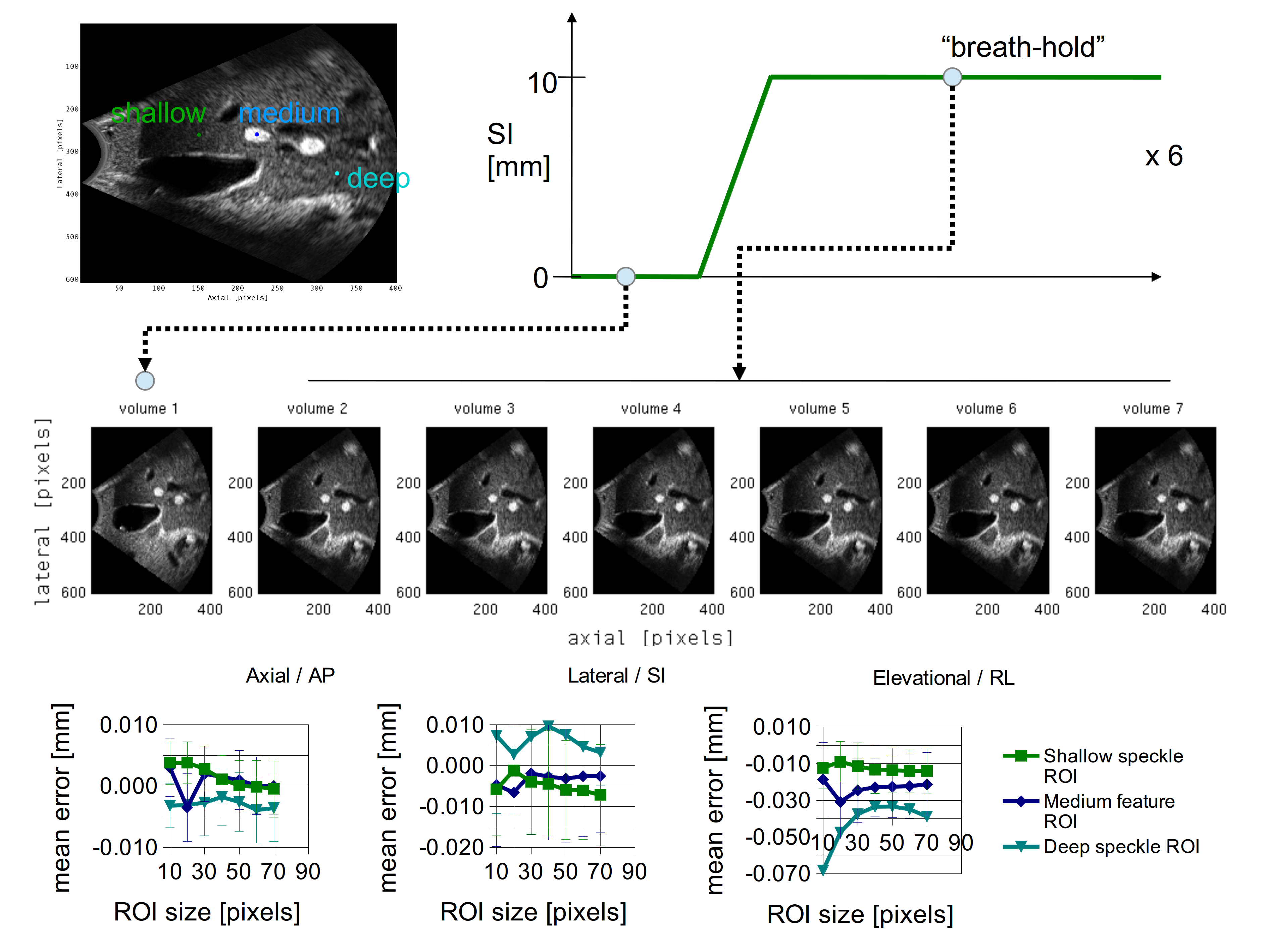
Figure 2. Reproducibility of simulated breath-hold (mean difference of 3D-US compared to ground truth (no motion) for repeated breath-hold volumes) as monitored by 3D ultrasound for three different region-of-interests.
Breath-hold monitoring
Arresting motion is a common method used to improve radiation beam delivery accuracy and reduce margins for treatment sites influenced by respiratory motion. A study is under way to investigate the use of ultrasound to monitor inspiration breath-hold as performed by the Active Breathing Coordinator™ (ABC) device. This work is being performed in collaboration with Johns Hopkins University (USA).