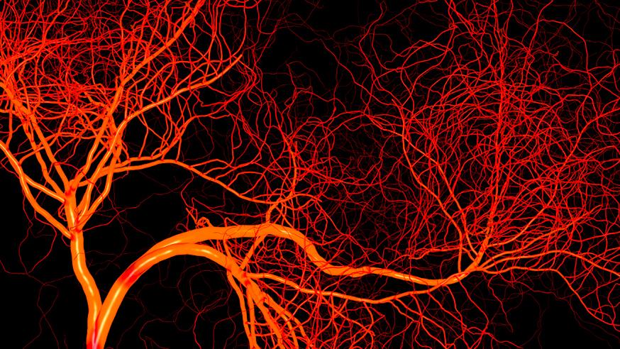
A magnetic resonance imaging test that measures the volume of blood in tumours could help select which patients will benefit from drugs that block blood vessel growth, a new study shows.
In a study published in the journal Cancer Research, researchers have also provided intriguing new clues about how cancers become resistant to such drugs.
Scientists at The Institute of Cancer Research investigated a type of magnetic resonance imaging (MRI) technology called susceptibility contrast MRI to measure blood volume in renal cell carcinomas – a type of kidney cancer.
In the study, funded by Cancer Research UK, the team used the MRI scanning method to quantify the blood volume of kidney tumours transplanted in mice, before and after treatment with sunitinib – a drug that blocks blood vessel growth.
They found that the drug caused the volume of blood in tumours to drop by an average of 70%, while their overall size remained the same.
The drop in tumour blood volume caused by sunitinib was linked with a decrease in the number of blood vessels in the tumours.
Tumours with the largest volume of blood to start with had the greatest drop in blood volume and the best response to treatment with sunitinib, the study showed.
Drugs that block blood vessel growth in tumours
The findings point to quantitation of blood volume as a new, non-invasive measure of response to sunitinib – and possibly also other drugs that block blood vessel growth in tumours.
In the future, measuring tumour blood volume could help identify patients with kidney cancer who would be most likely to benefit from treatment with sunitinib.
Intriguingly, the researchers also found that blood volume remained suppressed in tumours that had stopped responding to sunitinib, and vessels did not grow back.
This suggests that in sunitinib-resistant tumours, other mechanisms aside from new blood vessel growth are at work to allow the tumour to continue growing.
These mechanisms could include altered metabolism in tumours in the absence of oxygen – but need to be further studied, the researchers say.
Dr Simon Robinson's Magnetic Resonance team develops and tests new probes, instrumentation and techniques to better plan and assess cancer treatment.
Read more
Predicting which patients might benefit from sunitinib
Study leader Dr Simon Robinson, Team Leader in Magnetic Resonance at the ICR, said:
“We found that application of a non-invasive magnetic resonance scanning technique, known as susceptibility contrast MRI, could reliably measure changes in tumour blood volume after treatment with sunitinib.
“Scanning mice before and after treatment with sunitinib showed a decrease in tumour blood volume, which was most pronounced in those tumours with the highest blood volume before treatment.
“We showed that our test for measuring tumour blood volume could in future be used to reliably predict which patients with renal cell carcinoma might benefit from sunitinib – and also provides insight as to why some tumours become resistant to this drug.”